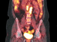
Functional Imaging

One of the main challenges in planning radiation treatments is that tumors aren't solid.
Typically — when cancer grows — it mixes with healthy tissue. This makes it hard to see where the tumor ends.

Doctors usually treat the entire area around the tumor. This approach targets the cancer, but also may affect healthy tissue nearby.
UPMC Hillman Cancer Center is one of the national leaders in using technology to accurately image cancers, and applying that data to plan the best treatments possible.
What Is Functional Imaging?
Most imaging methods focus on determining the location of a tumor in the body.
Functional imaging is the science of capturing what a tumor is doing, not just its physical location.
Radiation oncologists at UPMC Hillman Cancer Center devote much research to the design of many functional imaging methods, such as:
- fMRI (functional magnetic resonance imaging).
- MRSI (magnetic resonance spectroscopic imaging).
- PET (positron emission tomography).
- SPECT (single-photon emission computed tomography).
What Are the Benefits of Functional Imaging?
Functional imaging combines PET imaging with anatomic imaging methods — such as CT or MRI — to get a complete picture of a tumor’s:
- Composition
- Location
- Shape
- Size
The MRI or CT shows the physical shape and position of the tumor, while the PET scan shows the tumor’s activity.
Doctors use this information to plan your treatment.
Functional imaging gives doctors a clear picture of healthy tissue. This lets them use higher levels of radiation to treat the more active areas of the tumor, while helping spare healthy tissue and improve patient outcomes.
Contact Us About Radiation Oncology at UPMC Hillman Cancer Center
To learn more about radiation treatments or to make an appointment you can:
- Call 412-647-2811
- Contact a UPMC Hillman Cancer Center near you.

